Anterior Neck Anatomy Diagram / Root Of The Neck Last S Anatomy Regional And Applied : Many muscles are located in the anterior triangle of the neck.
byAdmin-
0
Anterior Neck Anatomy Diagram / Root Of The Neck Last S Anatomy Regional And Applied : Many muscles are located in the anterior triangle of the neck.. The boundaries of the intramuscular region of the neck are: The action of this muscle is lateral flexion and rotates the head to the opposite direction in unilateral contraction. Human_anatomy_skull_diagram 3/16 human anatomy skull diagram anomalies of the spinal cord. The final chapter deals with several diagrams showing the radiographs of the larynx, the skull, as well as the ventricular system of the brain. The two primary neck regions are the anterior cervical and posterior cervical triangles, which are found deep to the skin and subcutaneous tissue and contain several muscles, vasculature, and nerves.
The anterior triangle of the neck is made by the anterior border of the sternocleidomastoid muscle, the inferior border of the mandible and the midline of the neck. Browse 3,107 anatomy of neck and shoulder stock photos and images available, or start a new search to explore more stock photos and images. The action of this muscle is lateral flexion and rotates the head to the opposite direction in unilateral contraction. The neck is the start of the spinal column and spinal cord. Anatomynote.com found anterior view of the neck region artery, vein and nerves diagram from plenty of anatomical pictures on the internet.
Front On View Of Deep Anterior Neck Muscles Massage Therapy Yoga Anatomy Sports Massage Therapy from i.pinimg.com The neck contains seven of. Veins and arteries of the neck 9 photos of the veins and arteries of the neck activate javascript arteries in the neck diagram common carotid artery branches external carotid artery function how many carotid arteries left common carotid artery function the left. Pain in a man's body pain in a man's body on a gray background. For more anatomy content please follow us and visit our website: You can click the image to magnify if you cannot see clearly. The shoulder joint is formed where the humerus (upper arm bone) fits into the scapula (shoulder blade), like a ball and socket. Über 7 millionen englischsprachige bücher. As with all regions of the body, your study should start out with a look at the living region being studied.
This book is a valuable resource for radiologists, physicians, surgeons, and internists.
For more anatomy content please follow us and visit our website: As with all regions of the body, your study should start out with a look at the living region being studied. Veins and arteries of the neck 9 photos of the veins and arteries of the neck activate javascript arteries in the neck diagram common carotid artery branches external carotid artery function how many carotid arteries left common carotid artery function the left. Standard anatomical terms of location are used to unambiguously describe the anatomy of animals, including humans.the terms, typically derived from latin or greek roots, describe something in its standard anatomical position.this position provides a definition of what is at the front (anterior), behind (posterior) and so on. The posterior triangle of the neck is covered by the investing layer of fascia, and the floor is formed by the prevertebral fascia (see fascial layers of the neck). This triangle can be further divided into the submandibular triangle, submental triangle, muscular triangle and carotid triangle. The boundaries of the intramuscular region of the neck are: The cervical spine is delicate—housing the spinal cord that sends messages from the brain to control all aspects of the body—while also remarkably strong and flexible, allowing the neck to move. The anterior triangle is a region located at the front of the neck. The action of this muscle is lateral flexion and rotates the head to the opposite direction in unilateral contraction. Anatomy of the human pancreas explained with labeled diagrams. It is important to note that all triangles mentioned here are paired; The neck contains seven of.
The shoulder joint is formed where the humerus (upper arm bone) fits into the scapula (shoulder blade), like a ball and socket. It is important to note that all triangles mentioned here are paired; There are many muscles around the neck that help to support the cervical spine and allow you to move your head in different directions. We hope this picture anterior view of the neck region artery, vein and nerves diagram can help you study and research. Standard anatomical terms of location are used to unambiguously describe the anatomy of animals, including humans.the terms, typically derived from latin or greek roots, describe something in its standard anatomical position.this position provides a definition of what is at the front (anterior), behind (posterior) and so on.
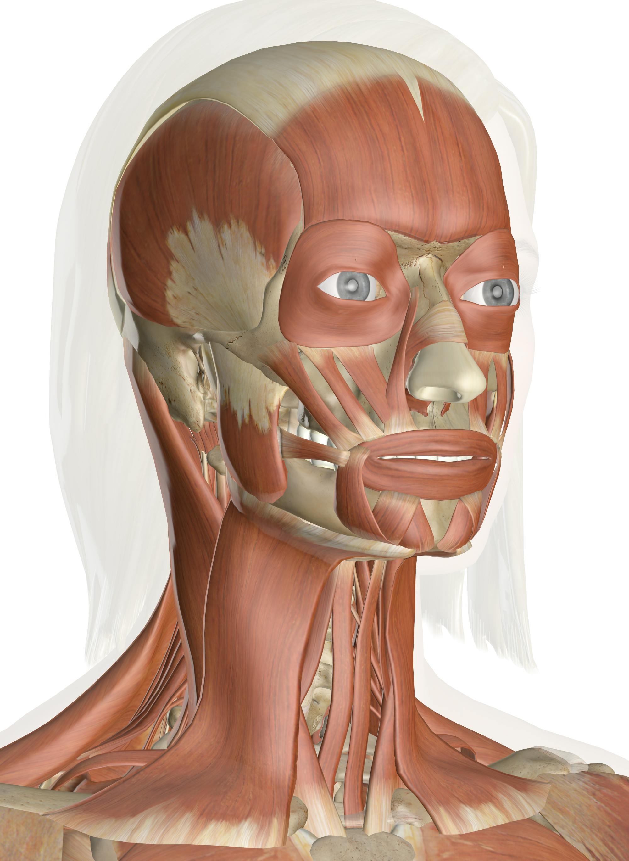
Pin On Career from i.pinimg.com The two primary neck regions are the anterior cervical and posterior cervical triangles, which are found deep to the skin and subcutaneous tissue and contain several muscles, vasculature, and nerves. They are located on both the left and the right sides of the neck. Muscles of the neck (musculi cervicales) the muscles of the neck are muscles that cover the area of the neck hese muscles are mainly responsible for the movement of the head in all directions they consist of 3 main groups of muscles: The boundaries of the intramuscular region of the neck are: Medially sagittal line down the midline of the neck. This book is a valuable resource for radiologists, physicians, surgeons, and internists. The neck is the start of the spinal column and spinal cord. The cervical spine is the most superior portion of the.
We hope this picture anterior view of the neck region artery, vein and nerves diagram can help you study and research.
It can be divided into 4 parts―the head, neck, body, and the tail. The pancreas, that somewhat look like the head of a golf club, does the job of producing digestive juices. Anatomynote.com found anterior view of the neck region artery, vein and nerves diagram from plenty of anatomical pictures on the internet. Standard anatomical terms of location are used to unambiguously describe the anatomy of animals, including humans.the terms, typically derived from latin or greek roots, describe something in its standard anatomical position.this position provides a definition of what is at the front (anterior), behind (posterior) and so on. This triangle can be further divided into the submandibular triangle, submental triangle, muscular triangle and carotid triangle. The neck is divided into several regions, triangles, and zones to organize the complex anatomy of this area. The action of this muscle is lateral flexion and rotates the head to the opposite direction in unilateral contraction. The final chapter deals with several diagrams showing the radiographs of the larynx, the skull, as well as the ventricular system of the brain. Head and neck anatomy,muscles,blood supply diagram. Browse 3,107 anatomy of neck and shoulder stock photos and images available, or start a new search to explore more stock photos and images. Learn vocabulary, terms, and more with flashcards, games, and other study tools. The shoulder joint is formed where the humerus (upper arm bone) fits into the scapula (shoulder blade), like a ball and socket. For intramuscular (im) injections, it is important to understand the anatomy of the neck region in some detail.
Common carotid artery (cc) internal carotid artery (ic) external carotid artery (ec) The pancreas, that somewhat look like the head of a golf club, does the job of producing digestive juices. Human_anatomy_skull_diagram 3/16 human anatomy skull diagram anomalies of the spinal cord. Here is a list of the many muscles that exist in the neck. A drawing of this region appears below.
Muscles Of The Head And Neck Anatomy Pictures And Information from innerbody.imgix.net The posterior triangle of the neck is covered by the investing layer of fascia, and the floor is formed by the prevertebral fascia (see fascial layers of the neck). The vertebrae (neck bones) running down and back from the back of the head to the point of the shoulder. The head of the humerus usually tears the inferior part of the joint capsule because this region is the least protected part of the capsule. The action of this muscle is lateral flexion and rotates the head to the opposite direction in unilateral contraction. The cervical spine's major functions include supporting and cushioning loads to the head/neck while allowing for rotation, and protecting the spinal cord extending from the brain. The anterior cervical lymph nodes are further down the front of the neck, divided into prelaryngeal, thyroid, pretracheal, and paratracheal, based on their position near structures of the throat. The head of the humerus usually tears the inferior part of the joint capsule because this region is the least protected part of the capsule. Muscles of the neck (musculi cervicales) the muscles of the neck are muscles that cover the area of the neck hese muscles are mainly responsible for the movement of the head in all directions they consist of 3 main groups of muscles:
Start studying head & neck anatomy.
Anatomy of the human pancreas explained with labeled diagrams. An understanding of this anatomy is essential for assessment and treatment of cervical spine problems. A drawing of this region appears below. The shoulder joint is formed where the humerus (upper arm bone) fits into the scapula (shoulder blade), like a ball and socket. Head and neck anatomy,muscles,blood supply diagram. The anterior triangle of the neck is made by the anterior border of the sternocleidomastoid muscle, the inferior border of the mandible and the midline of the neck. Veins and arteries of the neck 9 photos of the veins and arteries of the neck activate javascript arteries in the neck diagram common carotid artery branches external carotid artery function how many carotid arteries left common carotid artery function the left. Here is a list of the many muscles that exist in the neck. Standard anatomical terms of location are used to unambiguously describe the anatomy of animals, including humans.the terms, typically derived from latin or greek roots, describe something in its standard anatomical position.this position provides a definition of what is at the front (anterior), behind (posterior) and so on. Bodytomy elaborates more on the anatomy of the human pancreas. Anterior view of internal anatomy of neck region hyoid bone trachea and thyroid. The action of this muscle is lateral flexion and rotates the head to the opposite direction in unilateral contraction. The vertebrae (neck bones) running down and back from the back of the head to the point of the shoulder.